
Page 15 - 2016 July 專刊-H-0722
P. 15
【 In Situ Cell Death Detection Kit, AP / POD 】 ( Indirect TUNEL assay)
ROC-11684800010 / ROC-11684817910
偵測原理與標的 偵測
先使用帶有螢光分子之 Fuorescein-dUTP 去
標定細胞斷裂 DNA 之 3’OH。再利用帶有 AP
or POD 之 anti-fluorescein 抗體;專一性的 DNA
的斷裂
結合到螢光分子;之後加入 AP or POD 得進
行成色反應,即可進行影像觀察。
適用的實驗分析方法
一般的可見光顯微鏡
適用的偵測檢體
--細胞抹片
--組織切片(冷凍/石蠟包埋)
產品優點
• 可作單一顆細胞的分析
• 高敏感度低背景值
• 操作簡單,時間只需 3 小時
• 使用一般顯微鏡即可觀察
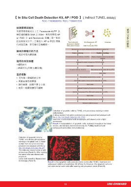
Detection of apoptotic cells by TUNEL and peroxidase staining in rabbit
endometrium.
A tissue section from rabbit endometrium was prepared and assayed with
the In Situ Cell Death Detection Kit, POD.
Slide was counterstained with hematoxylin and viewed under a light
microscope.
Result: A subpopulation of apoptotic cells, scattered throughout the tissue
section, are intensely stained (brown) by the TUNEL treatment and
subsequent peroxidase immunostaining.
Detection of apoptotic cells by
TUNEL and alkaline phosphatase
staining in rat spinal cord. A tissue
section from rat spinal cord was
prepared and assayed with the In
Situ Cell Death Detection Kit, AP.
The slide was viewed under a light
microscope (Panel A). After viewing,
the same slide was stained with
propidium
iodide and viewed by fluorescence
microscopy (Panel B).
Result: A few apoptotic cells (red) are clearly visible after TUNEL treatment and
subsequent alkaline phosphatase IHC (Panel A). However, the apoptotic cells are
not visible in the same slide after staining with propidium iodide (Panel B).
13 UNI-ONWARD