Page 7 - 2016 July 專刊-H-0722
P. 7

偵測粒線體膜電位改變


                                                                                                                     偵測粒線體膜電位
            【Mitochondrial Membrane Potential Kit】SI-MAK159 / SI-MAK160
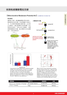
              偵測原理
               細胞凋亡初期,線粒體轉膜電位會發生變化,                        實驗操作流程
               導致膜穿透性的改變。利用是陽離子染劑JC-
               10,可以偵測到細胞膜的通透改變。JC-10在
               正常細胞中,會在線粒體中形成聚集體,發出
               強烈的紅色螢光。但是在凋亡的細胞中,因線
               粒體穿膜電位的改變,會以單體形式存在細胞
               液中,而發出綠色螢光。


                                                           Add 50ul of JC-10 dye ,
                                                           and incubate 30~60 min.
                                                           Then add 50 ul of assay
                                                           buffer B
                                                                                     Monitor the fluorescence
                                                                                     intensity (lex = 490/
                                                                                     lem = 525 nm) and (lex =
                                                                                     540/lem = 590 nm) for
                                                                                     ratio analysis. The ratio
                                                                                     of red/green fluorescence
                                                                                     intensity is used to
                                                                                     determine MMP.


                   Campotothecin-induced mitochondria membrane potential   Effect of FCCP induced mitochondria membrane
                   changes were measured with JC-10 and JC-1 in Jurkat   potential change in JurKat cells. JurKat cells were
                   cells. After Jurkat cells were treated with camptothecin   dye loaded with JC-10 dye-loading solution along
                   (10 μM) for 4 hours, JC-1 and JC-10 dye loading   with DMSO (Top) or 5 μM FCCP (Low) for 10
                   solutions were added to the wells and incubated for 30   minutes. The fluorescent intensities for both J-
                   minutes. The fluorescent intensities for both J-aggregates   aggregates and monomeric forms of JC-10 were
                   and monomeric forms of JC-1 and JC-10 were measured   measured with a flow cytometer using FL1 and FL2
                   at Ex/Em = 490/525 nm and 490/590 nm.           channels. Uncompensated data (left column) were
                                                                   compared with compensated data (right column).


                  相 關 產 品                          偵 測 方 式                 產 品 品 號            包 裝

                  Mitochondria Membrane Potential   Microplate Readers    SI-MAK159          500Test
                  Kit (JC-10 assay)                Flow                   SI-MAK160          100Test


                                                            5                                     UNI-ONWARD
   2   3   4   5   6   7   8   9   10   11   12