
Page 6 - 2016 July 專刊-H-0722
P. 6
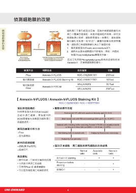
偵測細胞膜的改變 偵測粒線體膜電位改變
偵測細胞膜的改變
細胞凋亡不會引起發炎反應,因為在晚期細胞會形成 【Mitochondrial Membrane Potential Kit】SI-MAK159 / SI-MAK160
凋亡小體讓巨噬細胞、或是自噬細胞所吞食。研究發
現細胞凋亡時期,細胞膜會產生一些變化,這些細胞 偵測原理
膜的變化可能是一些“訊息”;讓體內啟動自我排除機 細胞凋亡初期,線粒體轉膜電位會發生變化, 實驗操作流程
制。細胞凋亡時細胞膜變化有以下幾個特徵 導致膜穿透性的改變。利用是陽離子染劑JC-
1. 膜表面糖蛋白的sialic acid residues消失 10,可以偵測到細胞膜的通透改變。JC-10在
2. 細胞失去原本細胞膜的不對稱性;例如:內膜的 正常細胞中,會在線粒體中形成聚集體,發出
物質Phosphatidylserine暴露至外膜。 強烈的紅色螢光。但是在凋亡的細胞中,因線
因此可利用對Phosphatidylserine具有高度親和性的 粒體穿膜電位的改變,會以單體形式存在細胞
Annexin-V,去偵測細胞膜的變化。 液中,而發出綠色螢光。
偵 測 方 法 相 關 產 品 產 品 編 號 包 裝
Flow Annexin-V-FLUOS ROC-11828681001 250Test Add 50ul of JC-10 dye ,
and incubate 30~60 min.
螢光顯微鏡 Annexin-V-FLUOS Staining Kit ROC-11858777001 50Test Then add 50 ul of assay
buffer B
Monitor the fluorescence
螢光顯微鏡 Annexin-V-FITC kit XR-LAVK050 50 Test intensity (lex = 490/
Flow XR-LAVK250 250Test lem = 525 nm) and (lex =
540/lem = 590 nm) for
ratio analysis. The ratio
of red/green fluorescence
intensity is used to
【 Annexin-V-FLUOS / Annexin-V-FLUOS Staining Kit 】 determine MMP.
ROC-11828681001/ ROC-11858777001
偵測原理與標的 實驗的操作流程
利用帶有螢光染料的AnnexinV、
去結合凋亡細胞;再搭配PI與
BOBI兩種螢光染劑區別細胞凋亡
與細胞壞死。
適用的實驗分析方法
--Flow
--螢光顯微鏡
Campotothecin-induced mitochondria membrane potential Effect of FCCP induced mitochondria membrane
changes were measured with JC-10 and JC-1 in Jurkat potential change in JurKat cells. JurKat cells were
適用的偵測檢體 cells. After Jurkat cells were treated with camptothecin dye loaded with JC-10 dye-loading solution along
--細胞(懸浮or貼附) 區別正常細胞,凋亡細胞與壞死細胞的染色結果 (10 μM) for 4 hours, JC-1 and JC-10 dye loading with DMSO (Top) or 5 μM FCCP (Low) for 10
minutes. The fluorescent intensities for both J-
solutions were added to the wells and incubated for 30
--組織切片 Normal Apoptotic Necrotic minutes. The fluorescent intensities for both J-aggregates aggregates and monomeric forms of JC-10 were
cells cells cells and monomeric forms of JC-1 and JC-10 were measured measured with a flow cytometer using FL1 and FL2
channels. Uncompensated data (left column) were
at Ex/Em = 490/525 nm and 490/590 nm.
產品優點 Annexin-V staining - + + compared with compensated data (right column).
• 操作快速:只需15分鐘染色時間
• 可快速分辨凋亡的細胞 Propidium iodide - - + 相 關 產 品 偵 測 方 式 產 品 品 號 包 裝
staining
• 可使用Flow 或 顯微鏡觀察
• 可以區別細胞凋亡或細胞壞死 BOBO-1 - - + Mitochondria Membrane Potential Microplate Readers SI-MAK159 500Test
Kit (JC-10 assay) Flow SI-MAK160 100Test
UNI-ONWARD 4