Page 17 - 2015 April 3D 細胞培養新趨勢
P. 17

3D CelluSponge 產品分類與適合的細胞株


                 產品               CelluSponge            CelluSponge-Galactose       CelluSponge-Collagen

                            HPC scaffold with no       Galactose conjugated HPC      Collagen conjugated
                 特性
                            bioligand conjugation      scaffold                      HPC scaffold
                            由羥丙基纖維素(HPC) 製             適用於需要半乳糖的細胞,                  適合需要膠原蛋白基質的
                            成,適用於不需要特定                 例如: 肝細胞在體外之培養,                細胞培養。例如: 幹細胞、                  3D Cell Culture
                            Ligand 的細胞,如一般細            藥物檢測與藥物開發中早期                  間質幹細胞(MSCs) 、神
              適合的用途
                            胞與癌細胞株。                    篩選使用。 播種1天後細胞                 經細胞培養。
                                                       形成3D功能性的球體,此球
                                                       體可保持穩定100天。

                            • Human breast cancer      • Primary hepatocytes (rat,   • Embryonic stem cells
                            cells (MCF-7)              human and monkey)             • Human mesenchymal
                            • Mouse embryonic          • Hepatic cell lines (Huh 7,   stem cells
              適合細胞株
                            fibroblasts (NIH-3T3)      Huh 7.5, HepG2 and hepatic    • iPS derived
                            • Human foreskin           progenitor)                   cardiomyocytes
                            fibroblast (HFF)                                         • Neuronal cells


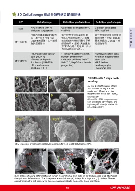
               A                             B                                NIH3T3 cells 5 days post-
                                                                              seeding

                                                                              (A) and B): SEM images of NIH-
                                                                              3T3 cultured on day 1 at low
                                                                              (scale bar 100 μm) and high
                                                                              magnification (scale bar 10 μm),
               C                              D                               respectively
                                                                              (C) and D): SEM images on day
                                                                              5 at low (scale bar 100 μm) and
                                                                              high magnification (scale bar 10
                                                                              μm), respectively.


              SEM images of primary rat hepatocyte spheroids formed in 3D CelluSponge-GAL.


              SEM images of neural differentiation of human mesenchymal stem cells on 3D CelluSponge-COL at different
              time points of differentiation. The time points are as follows: (A) 2 days (B) 7 days and (C) 14 days. The red
              arrow indicates the cell body, while the green arrows indicate the neurite. Scale bar 30 μm.


                                                           15                                     UNI-ONWARD
   12   13   14   15   16   17   18   19   20   21   22